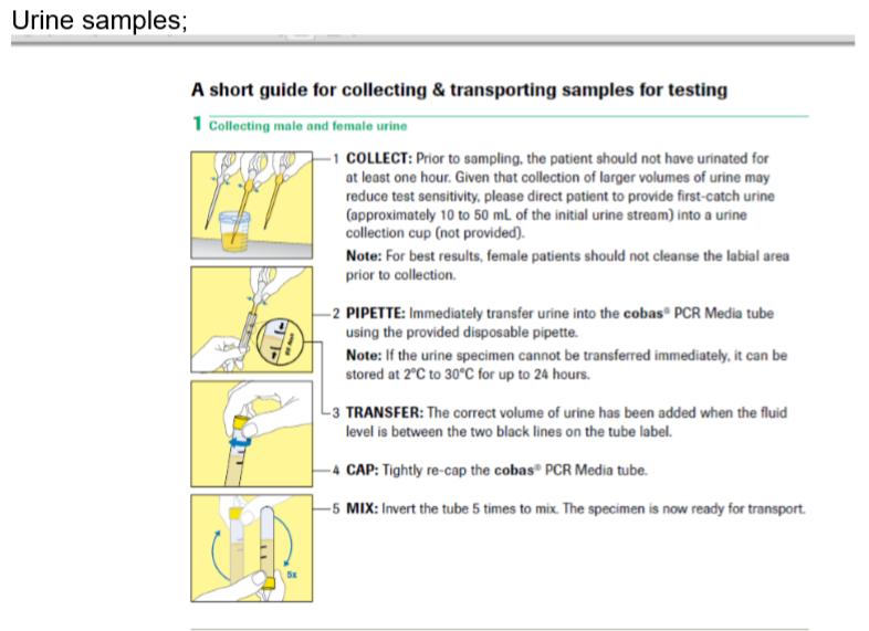
In asymptomatic women at low risk of syphilis or BBV who are attending for routine screening for STIs, examination is not necessary. Women can be given instructions for the self-taking of vulvo-vaginal swabs. Don’t assume that women will prefer not to be examined. Be aware that many women will want the reassurance of an examination, so unless she is in a drop-in clinic with no facilities for examination, she should be offered the option of an examination.
See instructions for self-taken vulvo-vaginal swabs
In all symptomatic women, contacts of STI or those at high risk of infection, an examination is indicated. Examination of the anogenital area should be performed with the woman in the semi-lithotomy position on a couch in a warm and well-lit room. See Box 3 for a summary. Examination may include:
Oral examination and sampling
Inspect the oral cavity and pharynx for lesions such as warts or ulcers, for example, those seen in secondary syphilis.
Take an oropharyngeal sample for dual NAAT testing for gonorrhoeae and C.trachomatis (this can be self taken if the patient prefers) in women who
- work in the sex industry
- report sexual assault
- are contacts of gonococcal infection, or
- have been diagnosed with gonorrhoea at an anogenital site (positive cervical, urethral, vulvovaginal or rectal GC NAAT test)
- have been presumptively diagnosed with gonorrhoea (GNID seen on microscopy of a Gram-stained smear of urethral or endocervical material)
Take an oropharyngeal sample for culture (in addition to the NAAT test) for gonorrhoea in women who are contacts of gonococcal infection, or have been diagnosed with urethral gonorrhoea at an anogenital site (see Gonorrhoea chapter for details of how to do this).
Notes
- It is only necessary to take samples for gonococcal culture in addition to NAAT tests in those who are being treated for possible gonorrhoea.
- NaSH requests: Chlamydia: pharynx, GC NAAT: pharynx, GC culture: pharynx
- Culture samples should not be self-taken and the sensitivity of culture is highly dependent on sampling technique.
- Pharyngeal testing for trachomatis is of unproven benefit. It is not possible to suppress the chlamydia result on the dual NAAT in use in our laboratory, so NAAT testing for N.gonorrhoeae inevitably involves testing for chlamydia too. There is no indication for routinely testing for pharyngeal chlamydial infection.
Genital examination and samples
- Inspect the abdomen, and palpate for tenderness, guarding and masses.
- Inspect the pubic area for pubis, warts and molluscum contagiosum.
- Palpate the inguinal lymph nodes, and note enlargement and tenderness such as in primary genital herpes.
- Inspect the labia majora for lesions such as warts or ulcers.
- Gently separate the labia minora from the labia majora. Examine the labia minora and, after wiping with a cotton wool ball (anterior to posterior or ‘front-to-back’ direction, inspect the introitus. Look particularly for warts and lesions of herpes (HSV).
Take a vulvovaginal swab for dual NAAT testing for N. gonorrhoea and C. trachomatis using the cotton wool-tipped plastic swab in the test collection kit, inserted about 5 cm into the vagina while pressing laterally against the vaginal wall and rotating gently with a swirling motion for a minimum of 10 seconds. Withdraw and break off the end into the buffer solution provided (this can be self taken if the patient prefers – see self taken swab instructions)

In women who are contacts of a partner with gonorrhoea:
- Obtain urethral material for Gram-smear microscopy by gently inserting a disposable plastic inoculating loop (10L) into the distal 2cm of the urethra, applying gentle lateral pressure and withdrawing.
- Culture urethral material obtained in the same way for gonorrhoeae (MNYC medium)
Pass a plastic vaginal speculum and examine the character of any vaginal discharge. A homogeneous milky-white discharge that coats the vaginal walls suggests bacterial vaginosis. Note inflammation of the vaginal walls, as may occur in trichomoniasis. See chapter on vaginal discharge.
In women who have reported a change in discharge, or where the discharge appears abnormal:
- Measure the pH using narrow-range pH paper, avoiding the alkaline cervical secretions. The paper can be applied directly to discharge at the introitus or a cotton wool tipped applicator stick can be used to sample discharge from the lateral or posterior fornix for application to the pH paper. In bacterial vaginosis, the pH of the discharge is >5.0
- Collect a sample of material from the posterior vaginal fornix, using a cotton wool-tipped applicator stick. Prepare a smear on a microscope slide for subsequent Gram-staining, and then suspend some of the material in a drop of isotonic saline on another slide.
- Examine (x40 objective lens: magnification x400:) the saline mount preparation for the motile trophozoites of Trichomonas vaginalis, fungal hyphae and “clue cells”.
- Examine (x100 oil-immersion lens, magnification x1,000) the Gram-stained smear for the spores and/or hyphae of candidal infection, the presence or absence of lactobacilli (in bacterial vaginosis, lactobacilli are reduced in number or absent, their place being taken by a mixture of Gram-negative and Gram-variable cocco-bacilli), and “clue cells”.
Note the appearance of the ectocervix and the character of any discharge from the endocervical canal; in chlamydial and gonococcal infections there may be a mucopurulent discharge.
In women who are contacts of a partner with gonorrhoea:
- Use a cotton wool-tipped applicator stick to collect material for microscopy for gonorrhoeae from the endocervical canal, smearing the sample on a plain microscopy slide.
- Culture cervical material obtained in the same way for gonorrhoeae (MNYC medium)
If there is pelvic pain, or other features suggestive of PID, perform a bimanual vaginal examination.
In women with symptoms suggestive of a urinary tract infection, haematuria or proteinuria. obtain a mid-stream specimen of urine for microscopy and culture
If there is risk of pregnancy, perform a pregnancy test on urine.
Perianal examination and anorectal sampling
Inspect the perineum, perianal region and anus for lesions such as warts.
Take a rectal sample for dual NAAT testing for gonorrhoea and C. trachomatis in women who:
- work in the sex industry
- report sexual assault
- are contacts of gonococcal infection, or
- have been diagnosed with gonorrhoea at an anogenital site (positive cervical, urethral, vulvovaginal or rectal GC NAAT test,
- have been presumptively diagnosed with gonorrhoea (GNID seen on microscopy of a Gram-stained smear of urethral or endocervical material)
Using the cotton wool-tipped plastic swab in the test collection kit, insert about 3 cm into the anal canal while pressing laterally against the rectal mucosa and rotating gently for a minimum of 10 seconds. Withdraw and break off the end into the buffer solution provided (this can be self taken if the patient prefers – see self taken swab instructions for MSM)
In women who are contacts of a male partner with gonorrhoea, who have a positive NAAT test for gonorrhoea at any site, or have a urethral or cervical slide positive for GNID, take a rectal culture for gonorrhoea. Use a cotton wool tipped swab inserted about 3 cm into the anal canal while pressing laterally against the rectal mucosa and rotating gently for a minimum of 10 seconds.
If there are anorectal symptoms, pass a proctoscope, examine the distal rectum and anal canal, and obtain the appropriate specimens for microbiological examination as described above.


